Services
Computed Tomography (CT)

Computed tomography scans, also called CAT or CT scans, are used to diagnose and evaluate a range of conditions, including detecting tumors, assessing injuries, and examining the chest, abdomen, and pelvis. During a CT-guided procedure our CT technologist directs a patient to lie on a table that moves through CT equipment in a machine called a gantry. The gantry houses an X-ray tube that rotates around the body and a computer processes this information through CT software to generate detailed cross-sectional images, or slices, of the internal structures.
The images produced by CT imaging and CT scans offer detailed information about the size, shape, and density of organs, tissues, and bones—providing valuable information in a CT report to help patients understand the full picture of their health.
What type of CT scans are offered at IMI?
A body CT scan involves getting cross-sectional images depicting bones, organs, soft tissues, and blood vessels. These images are then processed by a computer to construct highly detailed 3D representations. This imaging technique is key in locating, diagnosing and staging treatment for various diseases and disorders. It plays a crucial role in identifying conditions such as cancer, lymphoma, infections, heart and cardiovascular diseases, internal injuries or traumas, musculoskeletal issues, and a range of other medical concerns. The comprehensive insights provided by CT body scans contribute significantly to the accurate assessment and management of many other health conditions.
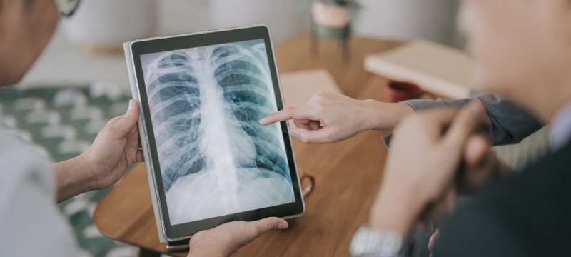
Low-dose Computed Tomography (LDCT) is a chest screening method with minimized radiation exposure, aiming to generate highly detailed images for the early detection of nodules and irregularities in the lungs. This rapid and painless non-invasive procedure involves capturing multiple lung images, enabling our radiologists to identify potential cancers before symptoms show up.
CT enterography is a diagnostic imaging examination employing CT scans to see the small intestine. This test is typically used to detect signs of inflammatory bowel disease, such as Crohn’s disease, as well as identifying gastrointestinal bleeding and small bowel tumors.
How to prepare for your CT visit.
Before your exam
- Change into the provided gown for the exam.
- If ordered, an intravenous contrast line will be placed in your arm.
- For abdomen or pelvis CT, you may have to drink oral contrast material.
During your exam
- Lie still on the imaging table for your exam.
- You may be asked to hold your breath during image acquisition.
For Small Bowel CT Imaging
- Have nothing by mouth (NPO) for at least 4 hours prior to the exam.
- Arrive at our Imaging Center about one hour prior to the examination time in order to drink a special oral contrast agent to distend the small bowel.

ACR Accredited in CT
IMI has been accredited by the American College of Radiology (ACR) for accuracy, safety and best practice standards in Computed Tomography (CT).